If the child does not see well you do not always see.
In this tab, I have summarized some of the information that I consider particularly important for new parents, knowledge of which allows you to detect and diagnose diseases in time even modest, but which could have a significant impact on the development of the vision of their children. It is essential for parents to consider the fact that their children are not able to evaluate a visual damage or serious vision loss (if the child does not see well do not always see), simply because they do not know what it means to see clearly, therefore difficult to, with the exception of serious diseases or for the appearance of a squint, small complain about a disturbance of vision. Also, and is most often the case, when one eye sees well and no other, the little patient may be apparently healthy parents because it can do everything with the eye normofunzionante. This condition can lead to a delay, sometimes years, visual identification of the problem and the correct therapeutic approach (Optical / ortottotica), resulting in the development of a lazy eye (ambliopa). This condition easily correctable in the pediatric age, becomes permanent adolescence and adulthood, without the possibility of recovery of a weak excellent vision in the eye that will remain so throughout life. For this reason, I recommend the reading of this sheet for parents and new mothers.
Alla nascita il nostro sistema visivo è perfettamente funzionante e quindi i bambini sono già capaci di percepire gli stimoli visivi, but their vision is limited to the perception of shapes and smoked lights, either because they are strongly hyperopic, both because do not have a good coordination of the eye muscles, but especially have not yet efficient cortical processing of images.
The first six months is critical for the development of motor and sensory function, that is completed around the 6-7 years with the full achievement of visual stability. The eye color e alcuni difetti di vista come la myopia, the hypermetropia , and l ' astigmatism , have a well-documented eredofamiliarità. These refractive defects, as well as other developmental changes in ocular, can lead to disharmony, delay or reduction of physiological visual acuity and must be recognized and treated early. Some eye diseases (p.e. congenital cataract, anomalie della parte anteriore e/o posteriore dell’occhio,etc) visual apparatus that occur in the first six months of life often involve, cause serious and irreversible vision. Damage to these characteristics that occur after the six months are responsible for a severe reduction in the infant's vision, but their timely treatment generally allows a good functional recovery.

The development of vision in children in looking at the mother. On the left (to 1 month) visual perception is limited to shades of gray and blurred; center: his vision to the 3rd-4th month, begins to perceive colors, recognizes faces and returns the smiles though still see a little’ blurred; right the sharp vision and color that is approximately 2 age.Mother's face. In the first month of life the child does not perceive colors again, then see in shades of gray, still unable to focus the images to approximately 20-30 cm, precisely the distance to learn to recognize the face of his mother when breastfeeding. In 3-4 month is able to recognize the human face answering smiles, grimaces and lip movements. And 'This is a very complex phenomenon that involves higher cortical structures (right hemisphere) and later with the feature of the temporal lobe cortex fails to recognize a face seen previously. This collaboration is essential to brain-eye and enables fast learning of the coordination of eye movements and the child begins to follow the moving image by rotating the head and have a certain degree of convergence if an object approaches the face. As mentioned, around 6 months now controls well the eye movements of convergence and tracking (disappears strabismus), able to have a sufficiently clear more than three meters and begins the "knowledge" of the world around him. The ability to distinguish the fundamental colors (red, green and blue) appears between the fourth and fifth months, while the stereoscopic sense (the ability to perceive images in three dimensions) begins to develop at the end of the first year of life to achieve full capacity, with subjective differences, after 2-4 age. The visual maturation continues until reaching a complete development around 2-3 years of life (ten / tenths, binocular vision, stereoscopic sense, ocular motility complete (ortoforica), although this period is extremely subjective and some processing power than can be completed later in the year.
The images of the fruits are perceived by emiretine Eye, transformed into nerve impulses here, travel through the optic nerves; cross (decussano) at the level of the optic chiasm that is continuous in the optic tracts to get to the lateral geniculate bodies. From here originates the optical radiation that leads to the occipital cortex, where the image is drawn primitively. The perfect collaboration of the eye muscles allows a perfect alignment of the eyes; in this way the images simultaneously stimulate symmetrical points of the two retinas (those corresponding retinal points = fovee). Under normal conditions the visual axes, that is, the line joining the object with the fovea, converge on a single point. The brain receives as a double image of each object, one from each eye, and has the ability to merge them into a single three-dimensional image. This ability is called fusion. The merger is possible only if the images sent to the brain come from corresponding retinal points of the two fovee are equal in size and sharpness. Between the two eyes, there is a certain distance that enables users to access the same object from two slightly different angles (then the two images fall on corresponding retinal points is not exactly). When this occurs the brain merges the images and also takes advantage of the slight difference in angle to reconstruct the exact location in space: in this way the brain is able to obtain a further information, that is, the three-dimensional perception. Is realized in such a way stereoscopic viewing of an object. This is normally done in the development and physiological cortical the image that is formed is stereoscopic and well-defined (Figure A). If, however, there is a defect of refraction (or other eye defects) that make "tarnished" the vision in one eye (figura B), the reconstructed image at the cortical level is due to the fusion of 1 sharp image and a blurry and the brain "penalize" in proportion to blur, the eye involved, up to provide a framework of “lazy eye”. If the tarnished image that reaches the occipital cortex is confusing to the brain, this may unknowingly “turn off” the information that comes with the eye refractive defect that does not sync with the other can become cross-eyed (Figure C).
How they form the view?
During embryonic development is mature the visual pathways, but the brain learns to "see" the images that come from the two eyes after birth. In fact, these images are processed (“fuse”) in a single three-dimensional image in the cortex brain (see Figure). This process is essential for a harmonious functional development of the visual system. For if a visual defect hidden or can not be evaluated by parents (see refractive errors), one of the two images coming from one of the two eyes is not clear and crisp as the one from the other, the brain receives two images no longer "stackable" and therefore is no longer able to "merge" into a single. In this conflict, the brain chooses to "turn off" the image less sharp and that comes more blurred. This unconscious selection involves a gradual and variable functional suppression of that eye, that can lead to a lack of development of stereopsi (that is, the ability to see in 3 dimensions) and a more or less severe vision loss in affected (amblyopic or lazy eye), to reach up to strabismus. As mentioned, this decreased vision, if it occurs in the first six months of life is often irreversible, while after this period is achieved a more or less deep degree of amblyopia. Before you realize that this problem will be faster and optimal recovery.
When you make your first eye exam?

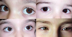
In the upper left, congenital glaucoma (buftalmo); in basso a sinistra una ptosi parziale congenita, to destra (above and below) esempi di congenital cataract.
While it is always carried out first eye examination at birth to all and to all premature infants at risk for genetically transmitted diseases before discharge from maternity wards, a complete eye examination is, in practice, assigned to the initiative of pediatricians and parents. As we have seen, it development of visual function occurs between the second and third year of age and it is therefore advisable to make at this time a first eye examination (except for rare conditions that require at a younger age, this is: congenital strabismus, leukocoria, buftalmo, etc for the meanings of these terms using “Search this site” in the upper right).With the frequency of Kindergarten, the teachers may notice abnormal visual behavior or eye movement disorder of the child that an added opportunity to detect possible refractive errors hitherto not highlighted. But the same parents can identify specific behaviors of their child who deserve to control eye, paying attention to any signs or attitudes, as for example: an abnormal position of the head (ie a particular inclination or rotation of the head that is not in straight, the squinting in the vision of far, the closure of an eye when fixing an object, it creasing frequent eye, increase in the frequency of closing of the eyelids (wink) tearing and redness. All conditions that require ophthalmologic evaluation.

Between the second and third years of age should make the 1st eye examination, because children are uncooperative and you can determine if any refractive errors, any disorder of eye movements, of stereopsis and fusion. Through schiascopia in cycloplegia can be determined refractive errors even if the child does not cooperate very. The eye examination must be completed, should any of the elements, visit the orthoptic (which consists in the accurate assessment of muscular balance of the eyes).
E 'then between the second and third year of life is recommended to take the 1st eye examination, because at this age is already possible to determine if present refraction defects, any disorder of eye movements, of stereopsis and fusion. Finally you can evaluate any ambliopie and the existence of a squint, with an excellent chance of recovery in a short time if you are working so early. The eye examination is especially important if the parents or pediatrician have found abnormalities in the eye and eye movements, or if you are in the family of hereditary eye diseases (is.: congenital cataract, congenital glaucoma, myopia, astigmatism, hyperopia.) Around 3 years it is possible to, as well as the eye examination, a full assessment of orthoptic (examination which assesses the state of the balance of eye movements), In fact, at this age the young patient is able to cooperate sufficiently and since the visit is that orthoptic eye represent a game for the child, is generally easy to obtain a valid collaborazione.Per its harmonic development of visual function there must always be a 'appropriate cooperation brain-eye, which can be considered sufficiently evolved around 6-7 years of age. So it is advisable to make a 2° eye examination at the time of going to school. A 6 years because the visual system is fully developed and can be assessed even modest refractive defects that can cause visual fatigue and mild ambliopie. School attendance is, for our baby, a new experience, where to learn many new things and where visual function is particularly useful (for example, learning to read involves fatigue and concentration not previously experienced by the child) and where you can highlight vision problems or ocular motility.

With an object that will draw the attention of our children we can evaluate the ability to follow eye movements in different parts of the visual field.
The eye examination in children begins with a discussion of problems reported by parents. The ophthalmologist should be informed of the general health condition of the child, if there were complications during pregnancy or at birth, if the growth and development shall regularly. The ophthalmologist will ask you if your child has had treatment with medicines, if it has undergone surgeries, If you suffer from allergies, if it has been subjected to special treatments eye, if you wear glasses, or if there are other disorders etc.. Continues with questions about the behavior of the small, if you take positions or unusual attitudes (abnormal position of the head, squint, tendency to approach to the TV, rub frequently eyelids, write in a distorted, school to judge the distance of the bench from the board, etc.), or investigate a reported symptoms by the child (fatigue in the reading, headache, burning eyes, clouding of vision), as well as aspects of the non-natural position of the eyes. It’ also useful to have a direct contact with the pediatrician, visiting the child more frequently than the ophthalmologist, so that such an exchange would highlight the onset pathologies.

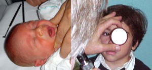
It is not difficult, intriguing little patient, see if it moves your eyes in every direction and converges well if approaching an object in the nose. (for example: in the picture in the middle you see the reflection "symmetry" of the light in their eyes).
It 's true, however, that parents, are very careful observation of their children and take their children to visit with very clear questions to ask your ophthalmologist. However, they may be useful in small test that they themselves can do with their children using common objects to the child and family (cars, colored pens, dolls, etc..). In addition, to keep his eyes, we can deduce its location capabilities and attention, if it moves at the same time both eyes in all positions and if this movement is harmonious or jerky. Giving up a curious object the child will closely converging eyes and you can evaluate this child's ability to improve after the first two months. With a little direct light on the eyes of the child to be able to assess pupillary function (ie if both pupils constrict) and the reflection of light on the eyes should be symmetrical (ie the light is reflected at the same point in both eyes).
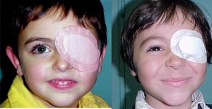
Still, with a subject that intrigues the child (or a play like in the picture below), one can try to cover one eye at a time of small and evaluate its reaction which must be equal to the occlusion of both eyes. Normally the small follow the object ignoring the occlusion, while if one eye sees less, occlusion of the best one will immediately attempt to remove the obstruction from the eye. The visual field child's age at birth is limited to central and then spread laterally to the first year of life, Therefore, always a subject that intrigues the small, one can follow the time evolution in its physiological side portions. It may for example, away more and more of the objects of interest to the small to see his vision, that is up to how far they can look up to and where to reach them. Finally, we must take into consideration the symptoms reported by young patients, symptoms related to an unconscious vice or defect of refraction (as the myopia, the hypermetropia , and l ' astigmatism), that may be responsible for visual fatigue (accommodative asthenopia) essentially characterized by headache, sometimes associated with burning eyes, frequent blinking and easy to read exhaustion.

With a dilated pupil and the eye to rest after the drops (cycloplegia) it is possible for the ophthalmologist locate a defect in the view of the child (schiascopia, left). The execution of a STEREOTEST (Titmus test) to destra, the child through the use of special glasses can see which of the images is rrilevata (three-dimensional) so you can study the degree of cerebral perfusion images of the two eyes and his sense of stereoscopic.
Contrary to popular belief, an ophthalmologist can realize if a child, very small, present visual problems based on the way down and follow objects, (index of the maturity of the visual pathways), as well as through special tests with the rotating disks, tables or instrumental. But even with the light, one can observe the reaction of the pupils, reflects the convergence, useful to see if the eyes see correctly, with the schiascopia, can be determined with good approximation the existence of a 'emmetropia (at this age the young patients are still sighted) and the presence of a vice or defect of refraction (as the myopia, the hypermetropia , and l ' astigmatism), and finally an examination of the ocular structures is possible to evaluate possible alterations of development.

On the left a optotype with “forks”, little patient indicates with his hands which side are the tips of the forks. In the middle child of 3 years without epicanthus and right same age with epicanthus. In the latter case, the parents mean that the child puts “the squint”. It is rather a pseudostrabismo determined precisely by epicantale fold more pronounced especially when the child looks to the side and looks like a squint.
There are also specific specialized tests that can be performed in selected cases. Older children, who do not know the numbers or the alphabet but speak, can reliably measure visual acuity, by special optotypes with symbols known to the world of childhood as birds, location, houses in decreasing size. It is important to examine the two eyes separately because very often the vision is different between one eye and the other. It should also assess the binocular vision and depth perception (stereopsi) by means of appropriate test. During this first part of the visit are also examined the attached external eye, such as the eyelids and lacrimal apparatus. By fixing the child with a portable lamp you get a bright reflection on the cornea which allows the ophthalmologist to check that the eyes are aligned. Exclude a squint is very important in young children because their nose, is not well-formed, can give a false impression that the eyes converge more than normal (escrow epicantale). During the visit the 'ophthalmologist covering first one eye and then the other: if the eyes are not aligned properly they will move in or out while fixing a light source or an object placed in front of (Cover test). A more accurate assessment of symmetry, convergence and coordination of the eyes occurs with the visit orthoptic. This particular visit is carried out by staff with a University Degree: The Orthoptist (assistant in ophthalmology), that assist the ophthalmologist in the research and treatment of any ambliopie, of eye movement disorder, imbalances sensory and ocular syndromes.

The pupil reacts to stimuli in normal light with a constriction (miosis left). The instillation of a few drops of collyrium based on derivatives of atropine, allows to obtain, in addition to the dilation of the pupil (dilated right) Also the block of accommodation of the eye (ie the possibility that they all eyes to vary its capacity to focus).
Why the eye doctor puts drops?
The part of the less grateful to the child, but indispensable, nell'instillazione consists of eye drops that allows the dilation of the pupils. Eye drops are administered one or more times and are effective, by 15-45 minutes, act by dilating the pupil and temporarily relaxing the power to focus the eye (accommodation). The tropicamide (Visumidriatic, Tropimil), has an effect of about 2-3 hours, while the cyclopentolate (Ciclolux), has a duration of approximately 7 hours. To have a greater efficacy in the block of 'accommodation it is necessary to instill the eye drops or ointment at home and complete at a later time the eye examination (the eye drops used in this case is l 'atropine whose effect on expansion lasts much longer, also 6-7 days and then you have to protect the little patient from the sun). With the partial blockage of accommodation you can highlight more precisely a visual defect (otherwise a control surface may also be fully offset by the high accommodative capacity of the small patient), accurately measuring the so-called “refractive defects” (myopia, hyperopia and astigmatism). And even in poorly cooperative children, it is possible to obtain data using refractive objective lens and a particular light source (retinoscope) through an examination which schiascopia. Projecting a beam of light into the eye, the ophthalmologist can evaluate, through the reflections of light and putting corrective lenses, if the child sees well (if it is cioèo defect of refraction (as the myopia, the hypermetropia , and l ' astigmatism),and then if you need glasses to correct in this way his visual defect. Furthermore with the dilation of pupil (midriasi) the ophthalmologist can examine the inside of the eye, ie the crystalline,, the retina, and the optic disc (the input of the optic nerve in the eye) to assess any developmental abnormalities or congenital.

Particular inclinations of the eye, the presence of epicanthus, characteristics conformations of the eyelids or eye rhyme may give the erroneous impression of a squint (pseudostrabismo).
Sometimes parents report that their children are "Squinty", but in reality the children, not yet having a good control of the eye muscles, soon get tired going generally in disagreement with the eyes. However, in the early 3-4 months there was a clear improvement because the ability to mature focus (accommodation), fixation and convergence and the child is able to perform movements rapid tracking with eyes. In fact, around the 6 this month pseudostrabismo disappears altogether. In some cases, however, (as in the picture) children seem to actually be crossed. This condition is caused by the fact that they have not yet well developed nasal and remains a certain degree of epicanthus which generally is reduced to around 6-7 age. However, it is always good to make an eye exam and possibly around the orthotic 3 years of age that can differentiate one by one pseudostrabismo strabismus real. In the latter case this is a condition in which the visual axes of the eyes are not parallel, but one eye can be deviated from inside or outside, upwards or downwards. Strabismus may be constant or intermittent. The children are usually unaware of their squint problem and this condition interferes with the development of the coordinated use of both eyes. Should therefore be treated with therapy orthoptic (bandage of an eye for a certain time), with the refractive therapy (glasses) the, sometimes if necessary, surgical (recession / resection of the eye muscles).
What are the defects dl view more’ common in childhood?

Graph survey on the conditions of eye health in kindergarten and the obligation to Rome (PDF file Meet clear ), the IAPB ITALY (International Agency for Prevention of Blindness – Italian Section), which shows that: the visit is made at the initiative of the parents in 83% of cases, and at the request of the pediatrician in the remaining 17%. Of the students who were taken by the ophthalmologist, the 26,9% wears glasses; compared to the whole sample (his 10825 cards), the percentage is 21,6% and that the error that is performed frequently is to delay the visit, which should be completed by the third year of life, in the mistaken belief that the older child is the more cooperative and more accurate assessments. However it appears that the 30% Children have never been seen by an ophthalmologist. With regard to the defect of view, myopia is reported in 33.02%, hyperopia in 21.55%, astigmatism in 26.79%, myopic astigmatism in 11% .89, the hyperopic astigmatism 6.74%. Visual defects are often not recognized in the age crucial for their correction and lazy or amblyopic eye may lead to possible evolution up to strabismus (eyes “crooked”)..
In general it can be said that the visual defect more frequent in this age group, but it is to be considered physiological factor associated with the growth of the eyeball is l 'hypermetropia . A 3 years, the refraction of children is on average +2,5 diopters (farsighted), a 6 years is approximately +1,50 diopters and a 15 years is situated around +0,75 diopters. Themyopia, grow significantly in the years following: juvenile myopia begins between 7 and 14 age (WGMPP, 1989) and increases with a rate of -0,40D / year (Goss and Cox,1985). However, the prevalence of myopia of at least 0,50 diopters varies greatly with the age of the population tested: a 5 years is lower than the 5%, reaches the 20-25% during adolescence and stabilizes in the adult as a percentage of 25-35%. (The prevalence of myopia in adulthood is still growing – 25% of myopia in the U.S. adult population (Sperduro,1983)). Theastigmatism, presents itself usually associated with one of the main defects, myopia and hyperopia, and is usually very stable during the life. It 'difficult to study the distribution of refractive defects in different populations and because they have a different geographical / ethnic (race, age, sex) and because in the various surveys the classification system can be different, as well as the methodology of measurement (visual acuity correct / incorrect, objective / subjective, ciclopegico / non ciclopegico). In addition, from birth to early 6-7 year 'sanatomy eye changes very (the axial length of the eye is at birth between 17.5 and the 18.5 mm and reaches towards the 13-14 years 24.00 mm), making it more difficult for a work of synthesis statistical. In a study of over 6000 children under the age of 2 years 1990 (PDF in lingua inglese Ocular and visual defects in a geographically defined population of 2-year-old children. M. Stayte, and to. ), ben il 2,1% of children had visual defects and emphasizes the relationship between these and an underweight at birth. About the 6 to the 9 % (Kendall e al. Ocular defects in children from birth to 6 years of age. Br Orthopt J 1989; 46:3-6) of patients in the visually impaired preschool, about 10% of school-age children with vision problems. In a recent study (2009) in Iran su 5913 children between 7 and 15 age (Prevalence of Refractive Errors in Primary School Children 7-15 Years of Qazvin City), has a prevalence of myopia 65.03%, hyperopia of 12.52%, l'astigmatism 16.1% and amblyopia of 6.36%. In another study (2005), Prof. F. Cruciani and Prof. A. Rinaldi publish an analysis of data on 10825 cards of a survey on eye health in kindergarten and compulsory, of Rome (file PDF Meet clear, the IAPB ITALY (International Agency for Prevention of Blindness – Italian Section)), which shows that: the visit is made at the initiative of the parents in 83% of cases, and at the request of the pediatrician in the remaining 17%. Of the students who were taken by the ophthalmologist, the 26,9% wears glasses; compared to the whole sample, the percentage is 21,6% and that the error that is performed frequently is to delay the visit, which should be completed by the third year of life, in the mistaken belief that the older child is the more cooperative and more accurate assessments. However it appears that the 30% Children have never been seen by an ophthalmologist. With regard to the defect of view, myopia is reported in 33.02%, hyperopia in 21.55%, astigmatism in 26.79%, myopic astigmatism in 11% .89, the hyperopic astigmatism 6.74%. Finally, as regards the prevalence of 'amblyopic or lazy eye, in the sample of the study shows that the number of children who performed the 'occlusion: 471 with a percentage of 4.4%. This result is a witness once again to the high frequency of this defect invalidating the need of prophylactic interventions as early as possible
The visit orthoptic

Some “tools” used dall'ortottista. To the left of the test Wirt (three-dimensional tables for evaluating stereopsis) the center and right sinottoforo lights Worth
As part of the visit, the ophthalmologist after the assessment of’visual acuity and the refraction in cycloplegia, may require, especially in children, the evaluation orthoptist to study problems related to the muscular balance of the eyes as the search for any ambliopie, of eye movement disorder, of strabismus and finally sensory imbalances. Theorthoptist (from the greek orthos = right = optein see), by special examination is in fact able to determine any functional alterations of the balance of eye movements. The orthoptist then run some tests as:
the cover test (CT). Through the occlusion direct (cover) od alternata (cover and cover) Eye compared to a target of fixation (for both distance and for near), allows to evaluate the presence or absence of strabismus or imbalances sensory minor (esoforie-tropie/exoforie-tropie, for meanings see "Search Site")
the ocular motility. We observe the ocular motility in both eyes in the various positions of gaze (9 positions) by looking at the patient a target-setting in order to identify possible impairment of synchrony and the mobility of the eyes.
stereopsis. (Test Lang, test di host): by means of special boards and sometimes with the use of polarized glasses, the patient should identify and recognize certain patterns that are raised (in 3 size "as if they were real"), respect to the bottom. This test provides information on the degree of cooperation of the eyes and the harmonious development of binocularity (In practice, if the patient uses both eyes simultaneously)
The test of Worth. Filters with red / green in front of the eyes observe the patient 4 colored lights for distance and near. If the patient has problems cortical fusion image can only see 2, 3 the 5 lights if there are problems suppression or diplopia
Examination sinottoforo. It 'sa particularly pleasing device to children because they see it as a game. E 'with two eyepieces through which the patient sees with two sheets of stickers and knobs by which you can move the images seen. Using this tool the orthoptist and able to "quantify the angle" of axis deviation Eye (if any strabismus or eteroforie), Furthermore it is possible to assess the degree of stereopsis, fusion and convergence. (Also with this tool you can assess even minor deficiencies of convergence and then use it in their correction by specific exercises).
To complete the diagnostic picture, for example those who have suffered trauma or diplopia post-traumatic orbital) the orthoptist can perform other special tests:
– testing with the aim bright red glass
– the visual field manual or computerized
– screen Hess-Lancaster
– electrophysiological examinations (pev,very,eog)
The Amblyopia
The improvement of visual function and the orderly development of ocular structures, its coordination to the eye movement in relation to the higher brain functions (maturation of the visual areas of the brain with representation "cortical" retinal), is realized, with some exceptions individual, around 2 years of age. If this improvement occurs before a change to the image formation on the retina (for the existence of refractive errors, damage to eyes, etc.) of an eye, the child begins to make more use of the other. The brain then tends to be used more and more in the vision the healthy eye resulting in poor exercise eye weaker, condition that involves, if not treated, a vision for making the condition more and reduced the so-called "lazy eye" (or amblyopic). Amblyopia, consists then, in a visual dysfunction in one or both eyes, structurally and clinically normal but have not had a functional physiological development. Amblyopia is istaura only in childhood, period during which it performs the development of visual function. If a visual defect occurs in the first six months of age is often irreversible, while after this period is achieved a more or less deep degree of amblyopia. If amblyopia is detected early (entered i 3-4 age) it is easier to ask remedy with great results; vice versa if the disease is diagnosed in late childhood, in the development of the brain dedicated to vision function completed, such a defect is difficult to fix and can not be longer repairable for the rest of their lives. High defects of vision in both eyes are easily recognizable by their parents because the child shows clearly does not see clearly: does not follow with his eyes, not trying to pick up objects with hands, can not trace a visual stimulus (light, object, etc.). It 'much more difficult for parents to highlight a flaw monocular, because the child can behave normally through the eye that sees well (even if it does not develop the three-dimensional sense). When you consider that the 'Amblyopia affects approximately 3-4% children, in Italy it would be with some 15-20.000 year. The most common causes that lead to amblyopia are:
– the refractive errors (myopia, hyperopia, e l'astigmatism) responsible for the 'amblyopia anisometropica
– strabismus (squint amblyopia) that does not allow the achievement of the same images on the two “fovee”,
– any visual defect that prevents the formation of a clear image on the macula (is.: congenital cataract, severe ametropia, anatomical abnormalities of the eye, ptosi etc.), ()
If the disorder of view is present in late childhood (after 8-10 age), ie when the visual function has completed its development, the risk of amblyopia is much reduced.
Prevention of amblyopia
The only effective preventive measure of’Amblyopia is a thorough eye examination that should be performed:->within the first year of life if there is a family hereditary eye problems
-> a 2-3 years of age to assess the proper development of visual function
-> school age, ie towards the 5-6 age.

Rehabilitation amblyopic eye. The occlusion of the healthy eye with special adhesive bandages, or filters applied to the lens of, stimulates vision in the eye lazy. This stimulation can be obtained, without bandage, also through good eye with atropine penalization, with the use of lenses or ipercorrette with filters to be applied on the lens of eyeglasses.
It l 'Amblyopia is determined by the presence of a visual disturbance to the load of an eye, his treatment consists of correcting refractive defects of the eye (with glasses), and in '"occlusion" healthy eye (with special adhesive bandages, or with filters on the lens of eyeglasses), in order to stimulate the visual function in the eye "lazy". As simple as this concept allows basic, under strict control eye within a short time, (even though the duration may be of years), to achieve amazing results with a visual and functional recovery almost always complete in affected. The variables that influence the outcome are many and different degree of impact (type of defect, magnitude of the refractive defect,age at diagnosis, maturation of the infant's vision and his collaboration, etc.). In this regard, it is important to clear determination and sacrifice some essential (Children will always try to remove the bandage or otherwise not to bring) by both parents, both on the part of young patients, continued use and rigorous treatment of the amblyopic (occlusion). If this fact, is carefully executed, you can get good results, also reducing the period of treatment. The therapy of amblyopia consists of two basic steps:
1) Resolution of the cause of amblyopia. The correction of refractive defects and anisometropie (different refraction in the two eyes) in children (with glasses or contact lenses after careful evaluation ophthalmology and orthoptics). The resolution of any factors of visual deprivation (this is. congenital cataract, ptosis, etc..) in cases where this is the cause. Strabismus trying to reduce as much as possible deviations, although this is often not sufficient to restore visual function. Congenital strabismus corrective action must be carried out as soon as possible
2) Rehabilitation amblyopic eye. We practice with the occlusion of the healthy eye with special adhesive bandages, or filters applied to the lens of, in order to stimulate the brain to develop visual function lazy eye. This correction can be obtained, without bandage, also through good eye with atropine penalization, with the use of lenses or ipercorrette with filters to be applied on the lens of eyeglasses. During the treatment the orthotic ocular conditions must be frequently monitored by the ophthalmologist and from 'orthoptist. E 'can also groped a direct stimulation of amblyopic eye (in the past experienced by Bangerter and Cuppers and called pleottica is now rarely used), through a tool that stimuli Pattern-flicker (STIMOLATORE PATTERN FLICKER MF17), can reactivate the visual lazy eye with good results in lighter forms of amblyopia.
Should also be considered that a significant visual impairment in a child in the pediatric age, well as compromising its visual function, may have an impact on its development with cognitive difficulties at school, learning and social relationships outside the family and family. This is a crucial moment for the future of the child view is appropriate and a great capacity for collaboration between the ophthalmologist, parents, the orthoptist and the small patient.
Lo Strabismo
When setting a target one eye is deviated from the visual axis of the other is talking about strabismus. Predisposing factors are certainly: inheritance, high refractive errors especially unilateral (as the myopia, the hypermetropia , and l ' astigmatism), the association with neuropsychological symptoms, some systemic syndromes. It also binds to eye diseases (glaucoma, cataract, ptosis, etc..), paresis of cerebral origin, paresis of one of the eye muscles.
On the basis of the deviation, which characterizes the "corner" of strabismus distinguish:
Strabismo paralyzed or incomitant(ie the angle of deviation varies in different directions of gaze). It 'secondary to paralysis or paresis of one of the muscles that move the eye and causes the appearance of an inability to direct the eye in the field of action of the affected muscle. For this reason, this strabismus, usually secondary to trauma or post-inflammatory processes (vascular, neurogenic paralisi III, IV and VI cranial nerve, paralysis miogene, myasthenia, distoridea myopathy and peripheral paralysis congenital (syndrome retraction of the eyeball and syndrome of the sheath of the superior oblique muscle), is more frequent in the adult. The symptoms depend on the underlying cause strabismus and ranges from simple abnormal position of the head with removal of the affected eye in infantile forms, to diplopia with dizziness to have occurred acutely (post-trauma or post-inflammatory). The treatment of the strabismus is essentially aimed at correcting the underlying cause, medical (if metabolic or inflammatory disease such as diabetes), with prismatic lenses to avoid diplopia and surgery in selected cases aimed at restoring means transposition of the eye muscles contraction in a certain field of action of the muscle paretic.
Strabismus concomitant or heterotropia (ie the angle of deviation of strabismus is equal in all directions of gaze) and are secondary to an imbalance sensorineural that adjusts the position of the eyes and their perfect motor coordination. In this type of strabismus, every muscle is functioning normally when studied individually, but if it is evaluated with both eyes open (ie the binocular vision), the weaker eye deflects squint. In this strabismus the deviation is always manifested in all positions of gaze and not be compensated by the person. Concomitant strabismus does not give any symptoms because it presents itself in an age of plastic vision that intervene effectively compensatory mechanisms (abolition, alternation) image from abnormal eye deviated (see drawings optic tract) which presents almost always a certain degree of Amblyopia and therefore the therapy will be targeted to the correction of both defects.
In latent squint or heterophoria (is esoforia = inward, both exoforia = outwards), deviation is kept "hidden" from the cerebral perfusion (see "how it develops the view") and strabismus only becomes apparent when it is lost (see: visit orthoptic / cover test) or when the vacancy concentration neuropsychic that the subject does, even unconsciously, to keep it (fatigue, fever, trauma, etc). Symptoms related to the effort to keep the merger are eyestrain headache which may intensify in the near vision, abnormal position of the head sometimes diplopia.
Congenital strabismus. The angle of strabismus is very often associated with pronounced vertical deviations, may be present at birth or appears in childhood before the 6th month and surgical therapy is associated with that orthoptic for the degree of amblyopia that tends to be present in the eye deviated.
Accommodative strabismus. Occurs early (between 2-3 years of age), as a result of incorrect hypermetropia that alters the ratio convergence / accommodation. The hyperopic child tends to compensate for his lack accentuating the accommodation and the accommodation consequently causes excessive convergence over up to determine strabismus. The therapy is based on the correction of hypermetropia.
Sensory strabismus. Occurs as a result of a reduced vision in one eye and therefore to a lack of fusion of the images. The surgical correction is.
Strabismus fixed. Congenital ocular deviation of both eyes inside or outside.
Strabismus, also, can be early or late onset, constant or intermittent (the deviation is present only at certain times of the day), unilateral (always affects only one eye and) o salts ante (affects the two eyes alternately), cyclic, Small-Angle (microstrabismo) (for details of these forms see utility / dictionary).
Therapy. The squint is always an amblyopic eye and so it is important for parents to know that in the presence of a true squint their little therapy should be istaurata as early as possible and that the best results, start the necessary treatment (glasses, occlusion and possible surgical correction) Already a few months of life. The presence of a squint translates itself in a clinical neurosensory an imbalance of the visual system, however, and rehabilitation will be long and difficult and will have to resort to the help of which we have already talked about of amblyopia. To start the most appropriate therapy is essential to the correct diagnostic approach in the form of strabismus that results after a thorough eye examination in cycloplegia atropinica and orthoptic. For example, in the case of strabismus accommodative simple correction with glasses of the refractive defect allows us to resolve it, and surgical correction is contraindicated, while congenital strabismus surgery should be as early as possible to allow for a proper visual development, as well as for the aesthetic appearance. In paralytic strabismus of the adult who does not present the risk of amblyopia, diplopia is a major problem, but can be resolved in the majority of cases, with the use of prismatic lenses, with the use of botulinum toxin and optionally with surgery. The choice of surgical strabismus in aged 6-7 age, varies in relation to the type of strabismus, of refraction of vision, age of the patient and his response to treatment with glasses and occlusion, ultimately in relation to its visual balance sensorineural. In the surgical treatment of the patient squinting and there are no fixed rules that each patient is a unique case to be assessed individually. Accurate assessment of ocular motility, allows the identification of which muscles are hyperfunctioning and which ipofunzionanti and then of how to program the surgery. However, surgical methods are essentially two: a weakening of the muscles that work too (Recession), that the muscle is disconnected from its attachment eye and lost some millimeters , or reinforcement of those that are deficient (Resection), in this case the muscles are shortened by a few millimeters maintaining the same insertion or with their duplicatura which offers the advantage of being reversible. The use of adjustable sutures allows you to adjust the extraocular muscle surgery the day after the surgery with the patient awake and cooperative. This technique is now widely used, should be reserved mainly for cases of adult strabismus with diplopia and the reoperations in adults. In recent years, has been successfully used the Botulinum toxin injected in doses that adequate in ocular muscle causes a temporary paralysis. Initially used in congenital strabismus at an early age (6 months) with great success, is also used even in the adult acquired strabismus (dysthyroidism).
How should the glasses for the child?
For our small patient glasses are almost always an "object annoying" and lived, in most cases, as an object that restricts their freedom. You should also keep in mind that unlike the parents, responsible in most cases the choice of eyeglasses, rarely the child is interested in the aesthetic and trendy glasses. If so it is important that participates in the choice of glasses that best meets the, we must remember that the glasses, unlike the adult, not only serves to correct its refractive defect, but it is the "means essential" to its proper development of sight. Finally, we can not ignore some of the characteristics that make it suitable to the anatomy of the face (the anatomical relationships, interpupillary distance etc.. are very different from those of the adult), his school life and relationships with friends and to any sports activities. Let's look at:
The frame, children up to 6 year it is best to choose a frame made of lightweight, flexible and metal-free (especially if the kids will play sports) best soft plastic so as to enable a normal life with other children and not limit it in games. Should be a large field of view adhering well to the face, (perhaps with the application of a belt to bar), that maintains the well in position on the nose. Furthermore, the eyes should be well centered both horizontally and vertically, and coincide with the optical center of the lens. The frame is in fact not "slide forward" as such decentralization can generate prismatic effects relevant (so the higher the optical power of the lenses) increasing or decreasing the corrective power of the lens and reducing the field of view of the child. Should have the nose-pads and hypoallergenic poggiaorecchie (possibly silicone), to allow a good grip in children who have not yet developed the nasal (radice the naso), especially in smaller (under 5 age). The upper edge of the frame must reach (and in smaller, cover) the eyebrow because the world of children lived from bottom to top frames and small end to watch the baby above the lens. Finally, it must frequently be "readjusted" optician (each 3-4 months) for the deformations and stresses whose use by children may procure.
The lenses. Are preferable shatterproof lenses (plastic or polycarbonate), either because they are lighter and less dangerous for a young patient, both because they weigh much less than glass (this is. high hyperopia), particularly significant given up with the poor development of the root of the nose in children is also recommended UV protection. These lenses but they scratch more easily and therefore it is recommended anti-scratch treatment. The lenses of children should be cleaned every day.
The cost should be limited because a child glasses should be changed often, both for the rapid modification of the anatomical relationships due to growth (especially nose and cheeks that make the small frame), both for the possible progression of the defect both refractive and the greater degree of wear (practically useless anti-reflective coating).
For sports activities specifically(swimming, tennis, soccer, basket, etc), there are special silicone or plastic frames with shatterproof lenses and cinghiola the neck that your optician will advise. Also in these cases even in small patients with high visual defects, it is possible to resort to the use of contact lenses.Finally I would like to emphasize the importance increasingly important made from UV radiation to the eyes of children.
Today we all carry sunglasses, but often neglect this precaution for our children. Instead they are the ones most prone to damage from ultraviolet radiation (ranging from keratoconjunctivitis solar to the late cataracts and macular degeneration), because a certain physiological protection of the eye at this wavelength is developed with age. In addition, children generally spend more time outdoors in places that parents and (beach, sea, snow, pool etc.) which is higher than the reflection of UV rays and further increase the damage to the eyes of children. Protecting our children from UV rays is simple enough to wear a wide-brimmed hat and sunglasses with high UV protection that can be easily found by your optician.


